Though your ankle joint performs the job like a hinge, it is much important than being an ordinary hinge joint. Your ankle comprises of various significant structures. It is considered as a stable joint, as it has a distinct design. It is essential to have a stable ankle joint, as it will help in withstanding 1.5 times your total body weight while performing activities like walking and around eight times your body weight while running.
Regular ankle function is essential for smooth walking with a well-balanced and effortless gait. The ligaments, tendons, and muscles, which provide support to your ankle joint perform as a team while propelling your body. Some conditions like an injury or dislocation can hinder the regular working of your ankle, causing immense difficulty while performing activities with pain and inflammation.
Our guide will be helpful to understand:
- Which parts are in your ankle
- The way your ankle works
Significant Parts of the Ankle
The significant parts of your ankle can be segregated into various categories, such as:
- Muscles
- Blood vessels
- Bones and joints
- Nerves
- Tendons and ligaments
The topmost structure of your foot is known as the dorsal surface while the sole structure is called the plantar surface.
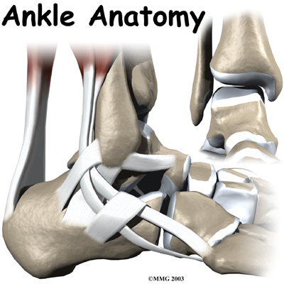
Bones and Joints
The joint of your ankle is connected by three bones. Your ankle bone is referred to as talus. The top part of this talus lies within a socket, which is a formation caused by your shinbone or tibia’s lower tip and a tiny bone at the lower end called the fibula. The lower part of your talus lies on the calcaneus or your heel bone.
Working of Talus Is Similar to a Hinge
The talus of your knee performs like a hinge within the socket to let you move your feet upwards and downwards. The upward movement is called dorsiflexion and the downward motion is known as plantarflexion. Carpenters and builders will be aware of your ankle joint’s design, as it is similar to the tools like tenon and mortise, which they use for creating beautiful structures with stability. They utilize it regularly for creating items with strength and sturdiness like buildings and furniture.
Tenon and Mortise
The bones present in your joints are wrapped using a slick substance called articular cartilage. This articular cartilage substance prevents friction and aids in smooth movement when moving the joints in your body.
Cartilage
The joints of your knee, hip, or ankle have a cartilage lining measuring the thickness of a quarter inch to help support your body weight. It is very soft and helps absorb shocks and sturdy enough to last until one’s lifetime, unless there is some kind of injury. The cartilage lining is present in between the tibia and talus to prevent the bones from rubbing against each other.
Tendons and Ligaments
Both tendons and ligaments are similar, which are actually smooth tissues, which help the attachment of a bone with the other. The only difference between them is that tendons perform the task of connecting the bones and muscles. These two structures are made with tiny fiber-like substance called collagen. These fibers of collaged join together like a bundle making it appear like a rope. Both tendons and ligaments are found in varying sizes and comprise of tiny fibers like that of a rope. The thickness of a tendon or ligament is directly proportional to its strength.
Collagen
Ligaments present in either side of the ankle joint help by holding the bones together. The lateral ligament complex comprises of triple ligaments located at your ankle’s farthest side, away from the other ankle. These three ligaments are called the PTFL or Posterior Talofibular Ligament, the CFL or Calcaneofibular Ligament, and the ATFL or Anterior Talofibular Ligament. Deltoid ligament is very thick that lies closely at the side of your other ankle to provide support to your medial ankle.
Three Major Ligaments
Ligaments provide support to your leg’s lower part where it acts like a hinge support for your ankle. Such ligament series provide ample support to your ankle syndesmosis, which is found at the tail end of the ankle paint where tibia and fibula meets. This part is supported by the tripe major ligaments. The AITFL or anterior inferior tibiofibular ligament lies just over the ankle’s front and connects the tibia and fibula. The posterior fibular ligaments like the transverse ligament and PITFL or posterior inferior tibiofibular ligament is found behind the ankle to attach tibia and fibula. Between the tibia and fibula, you can find the interosseous ligament, which is a connective tissue of massive length, which is present across the entire stretch of tibia and fibula from your knee towards the ankle.
Joint Capsule
The ligaments surrounding your ankle joint together form a part in the sac-like structure called the joint capsule. This joint capsule is a watertight structure resembling a sac that is present in every major joint in your body. It comprises of ligaments present around every joint as well as the soft tissues found amidst the ligaments, which fill up the gaps found in between them to form these joint capsule sacs.
Achilles Tendon
This largest tendon in your ankle plays a vital part in making you jump, walk, and run. It is connected with your heel bone or calcaneus and lets you stand on your toes. The calf muscles are connected by posterior tibial tendon from your foot.
Posterior Tibial Tendon
The posterior tibial tendon offers excellent arch support to your feet while the anterior tibial tendon lets you raise your feet. At the back of your lateral malleolus or ankle’s outer bump lies two tendons known as peroneals, which helps you turn the feet easily.
Ankle Muscles
Your ankle’s motion is supported by the strong muscles found in the lower leg with tendons connecting the foot and ankle. While walking, running, or jumping, these muscles contract to support the movement.
The functions of ankle muscles are discussed below.
- The peroneals, such as the peroneus brevis and peroneus longus lying at your ankle’s exterior edge helps in bending the ankle in and out.
- The calf muscles like soleus and gastrocnemius are connected with calcaneus by Achilles Tendon for bending the ankle by tightening the muscles.
- The posterior tibial muscles offers excellent arch support and aids in inward turning motion.
- The anterior tibial moves your ankle in upward direction.
Ankle Nerves
The nerves of your ankle travels till your foot to deliver muscle control and offers sensation to the outside edge and top part of your foot. The tibial nerve is placed just behind the medial malleolus.
Blood Vessels
The ankle receives blood from the arteries located close to it. at the top of the foot lies the dorsalis pedis, where you can feel the pulse.
Posterior Tibial Artery
The posterior tibial artery or the huge artery passed at the back of medial malleolus.
Summing Up
After reading this information resource, you will now be able to realize your ankle’s anatomy is complex. When all the structures and bones perform as a team, the ankle performs well. When a single part gets damaged, every part of your foot and ankle will be affected.